The +Ciencia magazine of the Faculty of Engineering shares interesting articles such as a chemiluminescence analyzer, the energy dispersion spectroscopy technique as an aid to better understand materials or the analysis of the Eiffel Tower model, as well as one on the electron microscope.
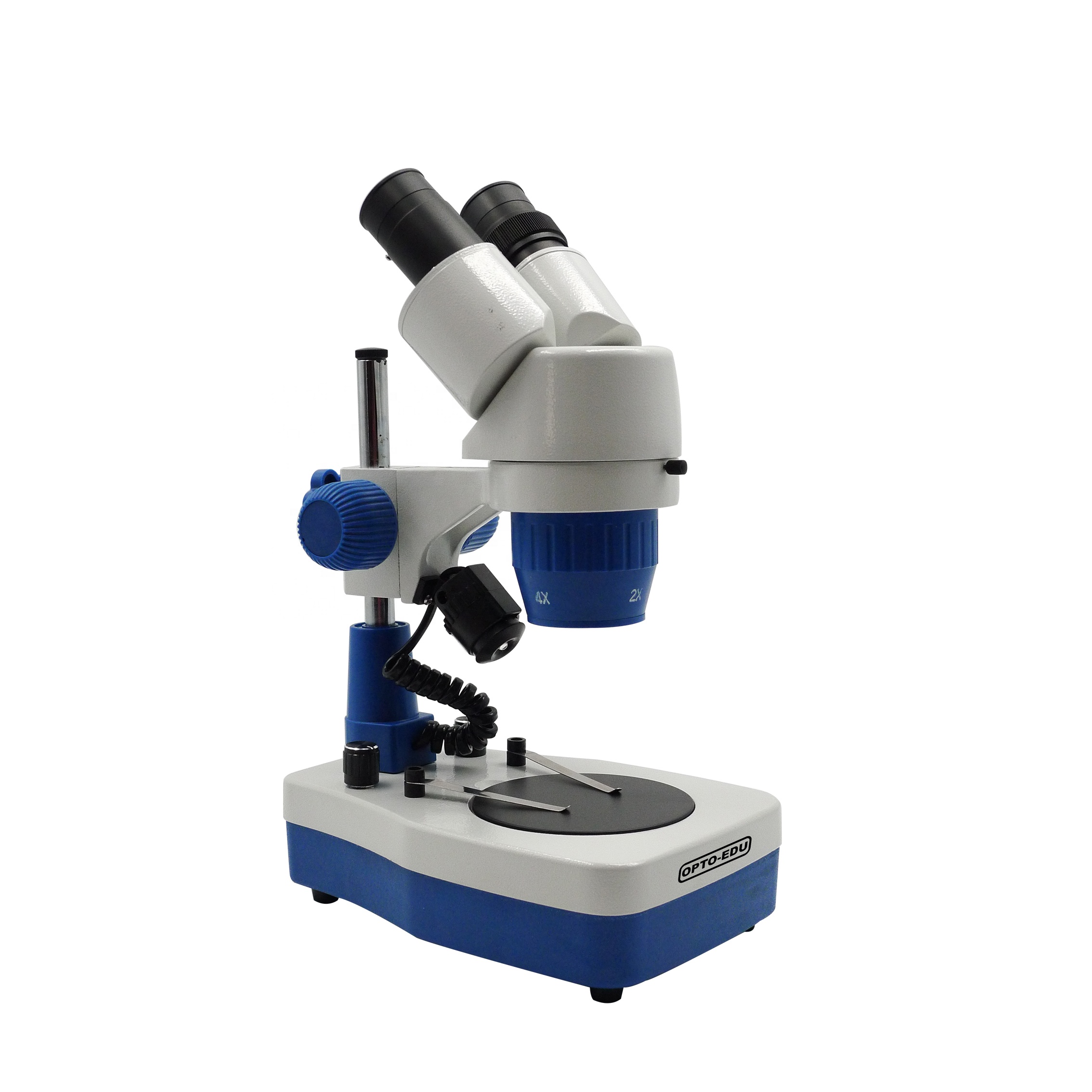
Since its invention, the electron microscope has been used in the study of the fine structure of living and non-living matter with a detail of dimensions of micrometers and nanometers, that is, for the study of ultrastructure. Labomed Microscope

Electron microscopes can be scanning or transmission.This work presents some applications of the transmission electron microscope in biological sciences, emphasizing its role in the study of cell structures that allowed the consolidation of cell biology.
Optical microscopes use light and glass lenses to generate images of objects with details down to 0.2 µm.By using electrons instead of light and electromagnetic lenses instead of glass, images can be obtained of objects whose details can be much smaller, down to about 0.005 µm.With the idea of overcoming the limitation imposed by the use of light as a source of energy in a microscope, the electron microscope was invented 90 years ago, in 1932. Thanks to its invention it was possible to go from analysis in the dimension of micrometers to the dimension of fractions of microns and even at the level of nanometers and angstroms.Over the years, the two types of electron microscopes that we know today were developed, namely: 1) the transmission electron microscope (TEM) and 2) the scanning electron microscope (SEM, for its acronym in English).The TEM allows us to study the internal structure of the samples, while the SEM allows the analysis of the surface structure.
The ability of the electron microscope to obtain a much higher resolution than that of the optical or light microscope favored its use in the study of the fine structure of matter, both non-living and living, in particular the study of the internal organization of the unit. structural of living beings, the cell.
First studies of biological material with TEM
Since the 17th century, both R. Hooke and A. van Leeuwenhoeck observed cells with the optical microscope.During the 19th century, T. Schwann and M. Schleiden postulated the Cellular Theory, proposing that all living beings are made up of cells.Indeed, this was the first robust theory that established biology as a science.This promoted the study of the structure of a large number of biological species with the optical microscope, strengthening that theory.However, due to the limitation of the resolution power of microscopes, it was not possible to advance in the knowledge of its fine structure that would help to understand its function in greater depth.Furthermore, the material had to be prepared to obtain very thin slices, a few microns or micrometers thick, through which the light radiating to the sample could be transmitted.
Due to the lack of color in biological samples, it was also necessary to develop staining techniques that would make it easier to distinguish between some components of the cell and others.Therefore, when the electron microscope is invented by Max Knoll and Ernst Ruska, the possibility of studying the cell in its fine internal structure opens up.Indeed, shortly after its invention in the early 1930s, Marton in Belgium prepared the first biological sample that he observed with the transmission electron microscope.
Professor Marton used a slice of a leaf of the carnivorous silver Drosera intermedia.Although he only observed with 65x magnification of the actual size, the resulting image showed a resolution or sharpness much higher than that achieved until then with the most powerful optical microscopes.In the image you can see a grid or sample support and a section of the leaf where epidermis cells can be seen.
First image of a biological sample recorded with the transmission electron microscope in 1932. It is the cut of a plant leaf placed on a metal mesh.
The use of the transmission electron microscope also allowed for the first time to observe the structure of viruses, until then only conceived as intangible pathogenic entities related to the word poison.One of the first observations was made by Professor Thomas Anderson in the early 1940s, with T4 bacteriophage viruses.The TEM images of the virus structure were so complex that they suggested they looked like tadpoles.Since then, transmission electron microscopy has facilitated the study of the morphological diversity of viruses.
Subsequently, studies of the structure of cells and tissues followed.Very soon it was noticed that, to study the internal structure of cells with TEM, it was necessary to carry out a sample preparation that 1) generated very thin cuts or slices through which electrons could be transmitted, that 2) eliminated remains of liquid, because the samples are introduced into a vacuum chamber and 3) were resistant to the impact of an accelerated electron beam.Additionally, biological samples contain mostly atoms of low atomic number, so they are almost transparent to electrons, which made it necessary to selectively add atoms of high atomic number, which would add additional contrast to the samples.Due to the above, several decades were required to ensure that the samples to be observed were cut into very thin slices, a few fractions of a micrometer thick—between 30 and 90 nanometers—that were stable to the impact of the electron beam and that all the liquid was extracted from them.
Thus, a standard procedure emerged for the preparation of biological samples for observation with the transmission electron microscope.Also, over time, variants of this standard procedure were designed, both for the observation of different types of samples and for the study of the composition and location of specific molecules.
Electron microscopy and cell biology
The use of a standard technique for transmission electron microscopy in biological sciences definitely contributed to understanding the fine structure of the cell.For example, the structure of the mitochondria was detailed and the presence of the so-called mitochondrial cristae, essential for understanding cellular respiration, was discovered.The rough endoplasmic reticulum and the Golgi apparatus, although they were first observed in 1902 and 1898 respectively, it was not until the 1950s that their existence was confirmed with the use of the transmission electron microscope.Likewise, thanks to the use of this instrument, the presence of vesicles and microvesicles related to the transport of molecules through the secretory route, of which these organelles are part, was confirmed.Likewise, it was possible to observe nanometric particles related to different metabolic events such as transcription, the respiratory chain or the morphological substrate of photosynthesis, i.e., the thylakoids of the chloroplast.
Likewise, the procedure for observing isolated structures such as ATP synthase, DNA and RNA or proteins.Notably, other discoveries such as the discovery of the ribosome by G. Palade and the endoplasmic reticulum by K. Porter, made with the transmission electron microscope, allowed the emergence of cell biology as a discipline aimed at studying the normal cell. and the pathological cell.
Lysosomes, microbodies or the fine structure of the nucleolus, the organelle where cytoplasmic ribosomes are produced in eukaryotes, essential for the production of proteins in all living beings, could also be described with this instrument.
Electron micrograph of a human cell.Different structures are observed in the nucleus (N) and the cytoplasm (C).In the cytoplasm the small arrows point to the mitochondria.The largest arrow points to the cytoskeleton.
Higher magnification ultrastructure of cytoplasm (C) of a human cell shows several organelles and submicroscopic structures such as the rough endoplasmic reticulum, which is indicated by the thin arrow.Mitochondria (thick arrow) and endosomes are also observed.(e) The cytoplasm contains abundant particles that correspond to ribosomes.
Transmission electron micrograph of a leaf cell of the teosinte plant (Zea perennis).The internal structure is observed, consisting of the nucleus (N) and nucleolus (nu), vacuole (V), mitochondria (m) and cell wall (Pc).The line indicates the 5 µm scale that allows the cell length to be calculated to about 20 µm.
Ultrastructure of a mitochondria (m).This organelle is covered by two membranes.The arrow points to a mitochondrial crista where the respiratory chain occurs.Outside this organelle there is rough endoplasmic reticulum (RER) and ribosomes (r) where protein synthesis takes place.
What do submicroscopic structures contain?
In addition to the study of cell and tissue structure, the production of molecular or chemical maps in situ has been an area of development.To this end, variants of the standard sample preparation procedure have been produced.Combined with the use of chemical markers or molecular probes such as antibodies and nucleic acid sequences, nucleic acid and protein localization techniques emerged that have contributed to the generation of molecular maps related to gene expression.Such is the case of immunoelectron microscopy and ultrastructural in situ hybridization of nucleic acids.With this, it is possible to know the in situ composition of submicroscopic cellular structures that contain genes and the products of their expression, such as nanoribonucleoproteins (nanoRNP).
New techniques such as electron cryomicroscopy already indicate a development in the knowledge of cellular structures at the level of molecular resolution within the cell, both prokaryotic and eukaryotic.Without a doubt, advances are aimed at knowledge at the molecular level of cellular structures, as well as their gene composition and expression products, which will allow knowledge of the specific functions in the life of cells.
Nowadays, the study of intracellular territories related to the organization of genomes and their products in the form of RNA and/or proteins are the subject of analysis.
We appreciate the technical support of Biologist Saraí Cruz Gómez and Biologist Diego García Dimas, for the preparation of the teosinte sample.
*Article by Luis Felipe Jiménez García and María de Lourdes Segura Valdez, researchers from the Department of Cellular Biology of the Faculty of Sciences of the National Autonomous University of Mexico (UNAM).
Referencias - Alberts, B., Johnson, A., Lewis, J., Morgan, D., Raff, M., Roberts, K., & Walter, P. (2015). Molecular biology of the cell, 6th ed. Garland Science. - Fawcett, D. W. (1981). The cell, 2nd ed. Saunders. - Marton, L. (1934). Electron microscopy of biological objects. Nature, 133, 911. - Pollard, T. D., Earnshaw, W. C., Lippincott- Schwartz, J.,& Johnson, G. T. (2017). Cell Biology, 3rd ed, Elsevier. - Porter, K. R., & Bonneville M. A. (1973). Fine structure of cells and tissues, 4th ed. Lea and Febiger.
Click on the cover to see the full magazine
For more interesting articles like this, we invite you to consult +Ciencia, the quarterly magazine of the Faculty of Engineering, which in each issue has relevant content from its students and researchers.Click here to see all editions.
More information: +Science.Magazine of the Faculty of Engineering Dr. María Elena Sánchez Vergara elena.sanchez@anahuac.mx
Av. Universidad Anáhuac 46, Col. Lomas Anáhuac Huixquilucan, State of Mexico, CP 52786. +52 (55) 5627 0210

A14 Inverted Av. de los Tanques no.865, Col. Torres de Potrero, Mexico City, Alcaldía Álvaro Obregón, México, CP 01840. +52 (55) 5628 8800